2 cases of female patients who suffered FOOSH type injuries involving the elbow. One (#06)caught her high heels on the carpet- walking.
No prior trauma, surgeries, carcinoma or corticosteroid regimen reported by either patient.
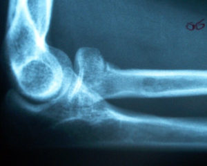
The radial necks shows a small cortical infraction. No abnormal fat pads were detected on the elbow radiographs.
If fracture lines are difficult to see, an additional Coyle view can be used.
This case shows cortical infraction with a vertical radiolucency extending inferiorly- at the radial neck.
Of incidental note, triceps muscle insertion site enthesopathic changes are present.
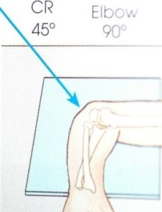
Coyle view (axial mediolateral ) view. Have patient sit a the edge of a table in a typical lateral view of the elbow. The hand is in pronation. The CR should enter the elbow joint at a 45 degree towards the humerus. (4)
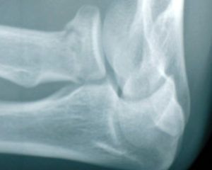
The Coyly view enhances the vertical radiolucency of the non-displaced radial neck fracture and removes the superimposition of the radial head from the capitellum.
DISCUSSION:
While fractures of the radial head may seen in all age groups, they usually occur in adults (85% between 20-60 years of age) and more frequently in women (M: F 1:2) (1).
These cases show the application of the Coyle view in fractur decision making. (2)
REFERENCES: