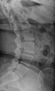
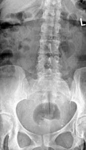
47 yo/female presents complaining of non-specific lower back discomfort. No prior red flags.
What do you see?
The AP view of the lumbar spine shows a curvi-linear density superimposed on the proximal sacrum. The latearal view shows the cyst/mass like density to be intra-peritoneal in location which measured 9.3 cm. in transverse dimension on initial x-ray interpretation. Mild spondylosis is present at L3 & 4. A diagnostic US of the pelvis was ordered.
TEACHING POINTS:
Uterine fibroids are synonymous with uterine leiomyomas or myomas.
Approximately 20-30 % of females of reproductive age will develop this mostcommon gynecologic tumor.
A patient with a 6.0 pound uterine fibroid (post-surgical specimen) is the author’s most vivid recollection of this condition!
The diagram below shows common locationsfor uterine based fibroids.
The new FIGO system divides uterine fibroids into 3 major categories based on their predominant location.
submocosal
other
hybrid
This recent system allows more uniformity in reporting and surgical planning. (1)
Ultrasound is the inital investigative modality.
MRI adds further evaluation of enlargement & assessment of submocosal lesions. This tool also helps characterize aggressive lesions from the more common fibroid.
COMMON LOCATIONS FOR UTERINE FIBROIDS.
REFERENCES:
Abdominal Radiology (2021)46:4146-2155 -MRI -based pictorial review of the FIGO classification system for uterine fibroids:
Case compliments of Dr.Permenter, DC Charlotte, North Carolina.
 What do you see?