Case 31- 74 yo/M patient Presents Left Neck Discomfort.
Case 31- 74 yo/M patient Presents Left Neck Discomfort.
What do you see?
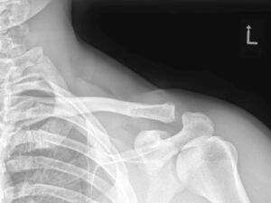
LEFT SHOULDER – IR VIEW
DISCUSSION:
The Rockwood classification system of AC joint injuries the most frequently used. Type I above is normal & Type VII is complete disruption of AD joint soft tissue holding elements including the coracobrachialis muscle.
REFERENCES:
1.Imaging of the AC Joint: Antanomy, Function, Pathologic Features & Treatment. RadioGraphics
https://doi.10.1148/rg.2020200039
2. Radiopadia.org
CERVIAL – AP-OM VIEW shows metallic surgical hardware superimposed on the maxilla region.
CERVICAL AP-LC VIEW -showing the surgical hardware previously mentioned.
Did you notice the sternotomy sutures and more importantly the vertical radiodensity to the left of the lower cervical vertebrae? This represents extensive left carotid artery calcification.
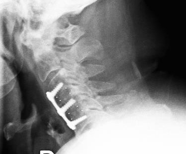
CERVICAL SPINE-Lateral CLOSE-UP VIEW-shows an anterior cervical dynamic plate C3-5 with small round radiodensities within the disc spaces. These are disc replacements.
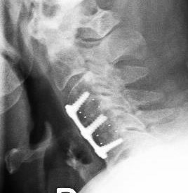
At C5, disc disease is present and an incidental C1 arcuate foramen is present. Nuchal ligament calcification is seen posteriorly.
CERVICAL SPINE- LATERAL-CLOSE-UP. The small metallic radiodensities represent disc struts of the disc replacement. The surgical arthrodesis involves the C3-5 motor units with disc replacement intervention of C3 & 4, therefore not a typical ACDF ( anterrior cervical dynamic fusion) but an anterior cervical disc arthroplasty (ACDA).
The extensive left carotid artery calcification – seen best on the AP-LC view warrants a diagnostic US of the carotid/vertebral artery tree.
Cervical disc disease affects up to 84 persons per 100,000 of the population. (population -based study -Rochester, Minnesota, Brain 1994. (1)
Traditionally, the ACDF was performed in patients with symptomatic single-level level disease. The procedure historically reduced local mobility however caused significant adjacent disc disease.
This case involves multiple level anterior cervical disc arthroplasty whether from autogenous graft ( iliac crest) or allogenic bone graft- cadaver or animal bone graft. (2)
The newer ACDA procedures involves various new artificial discs. The current literature is inconsistent with methods regarding clinical, radiological & patient outcome performance markers. (1)
Case 31- 74 yo/M patient Presents Left Neck Discomfort.
What do you see?
LEFT SHOULDER – IR VIEW
DISCUSSION:
The Rockwood classification system of AC joint injuries the most frequently used. Type I above is normal & Type VII is complete disruption of AD joint soft tissue holding elements including the coracobrachialis muscle.
REFERENCES:
1.Imaging of the AC Joint: Antanomy, Function, Pathologic Features & Treatment. RadioGraphics
 LEFT SHOULDER – IR VIEW
")
")