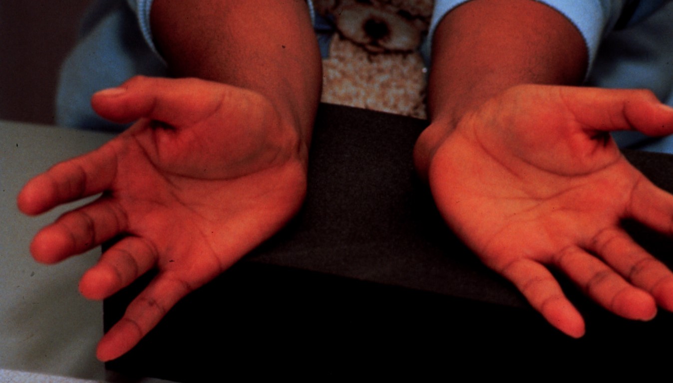
20 yo/F presents with left wrist discomfort ~ 12 months duration. Left ulnar N distribution discomfort that awakes her. No prior surgeries, carcinoma or corticosteroid regimen.
20 yo/F presents with left wrist discomfort ~ 12 months duration. Left ulnar N distribution discomfort that awakes her. No prior surgeries, carcinoma or corticosteroid regimen.

Nodular density- proximal to left hypothenar region.

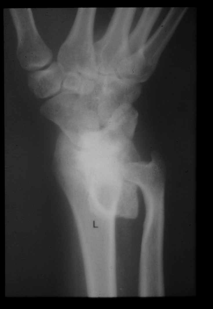
Semi-supinated view of the left wrist (Norgaard’s position)
Bone or soft tissue?
Questionable density seen on volar aspect with wrist with distal ulnar resorption.
Radiographic features: exostosis with chondroid matrix, parent
cortex continuous with lesion. All features consistent with
osteochondroma of volar left wrist.
Once the degree of encasement of the osteochondroma was verified with imaging, surgical intervention resulted in a successful clinical outcome.
DISCUSSION:
Osteochondroma represents the most common bone tumor is an aberration of enchondral ossification, NOT a true neoplasm. (1) Complications include: cosmetic and osseous deformity fracture, neurologic sequlae as in our case.
KEY FACTS: (2)
At ProImaging we provide expert chiropractic radiology interpretation.