60 yo/M patient presents with neck discomfort & LBP. No prior trauma, corticosteroid/opioId regimen or carcinoma is reported. AS has been diagnosed.
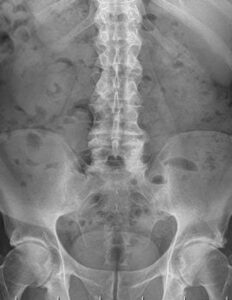
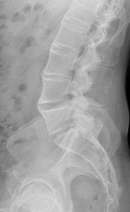
 AP & LATERAL VIEWS OF LUMBAR SPINE.

Non-visualizaton of the SI joints is the first clue that AS is present.
The lateral view shows the classic syndesmophyte formation, discal calcification & nonvisualization of the posterior lumbar joints.

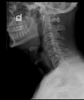
The lateral cervical spine views shows a small occipital exostosis, an arcuate foraman, spondylosis but importantly lower cervical syndesmophyte formation.
AS patient are at RISK for spinal fractures- 4x so. (1)These may subtle to detect & if SCI (spinal cord injury) is associated, a poor prognosis is associated. (2)
REFERENCES:
1.https://radiopaedia.org/articles/ankylosing-spondylitis-1?lang=us-AS
2.Spinal cord injury after traumatic spine fracture in patients with ankylosing spinal disorders
PMID: 28984512 DOI: 10.3171/2017.5.SPINE1722