Patient suffered prior FOOSH type injury.
No prior surgeries, carcinoma or corticosteroid/opioid regimen reported.
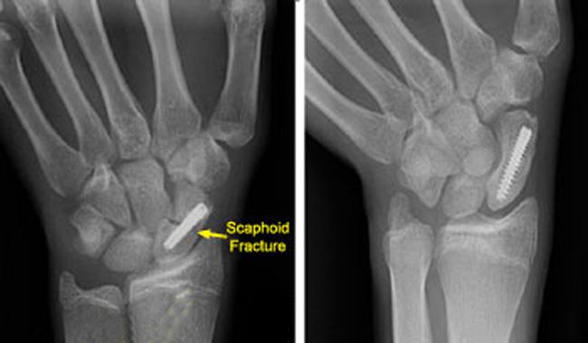
A cannulated screw transfixes the waist scaphoid fracture. This is a common treatment for internal fractures with > 1. 5 mm. displacment. (2)
As the majority of the scaphoid (8)%) is covered with articular cartilage, entry points for internal fixation are limited. (3)
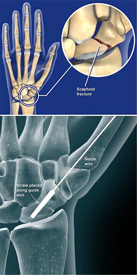
Wrist- anatomic model showing scaphoid fracture fixation.
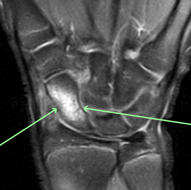
Coronal fat-sat MRI- arrows pointing to bone marrow edema and mid waist fracture line.
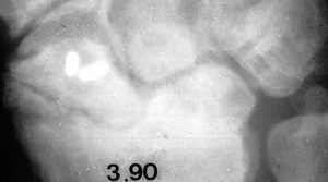
25 yo/M electrician showing mid waist fracture seen on older tomogram image.
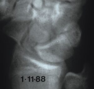
Follow up image – 2 years show advanced radial carpal collapse.
Early detection with MRI is essential for improved surgical outcomes.
DISCUSSION: After a FOOSH type injury, scaphoid fractures account for a large number of carpal fractures.
The majority are mid-waist fractures. If plain radiographs do NOT detect 5-20% of fracture, ulnar deviation views are helpful.
If clinical symptoms persist, the most sensitive modality-MRI-will detect subtle fractures and evaluate for osteonecrosis. (1)
REFERENCES: