Patient presents with left wrist discomfort after a FOOSH type injury.
No prior surgeries, carcinoma or corticosteroid/opioid regimen reported.
What do you see?
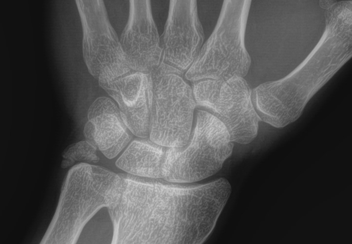
A semi-lunar density is seen where the normal left ulnar styloid process should normal be. Malunion or non-union of the ulnar styloid process is common after FOOSH injury. Associated triangular fibrocartilage injuries commonly associated.
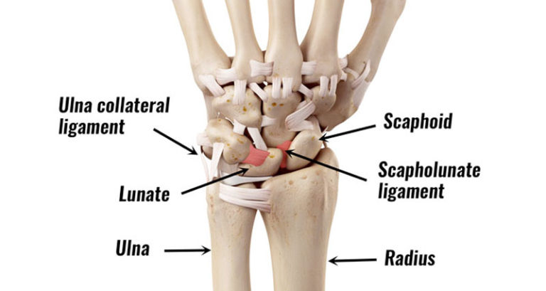
Wrist- anatomic model- dorsal perspective. The white horizontal density represents the triangular fibro-cartilage (TFC). (3)
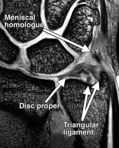
Coronal fat-sat MRI- arrows pointing to normal structures on ulnar side of wrist.
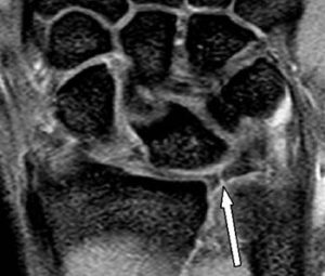
Coronal Fat-Sat image. The arrow demonstrates a complete tear of the radial side of the TFC. (1)
DISCUSSION:
TFC injuries are usually degenerative or post-traumatic. Normal variants in this region must be differentiated on MRI for accurate diagnosis. (2) Because MRI is invaluable in detecting & locating TFC tears, this helps in non-operative & operative decision making for the clinician. (1)
REFERENCES: