45 yo/M patient presents with “deep” LBP.
No prior trauma, surgeries, carcinoma or corticosteroid/opioid regimen reported.
What do you see?
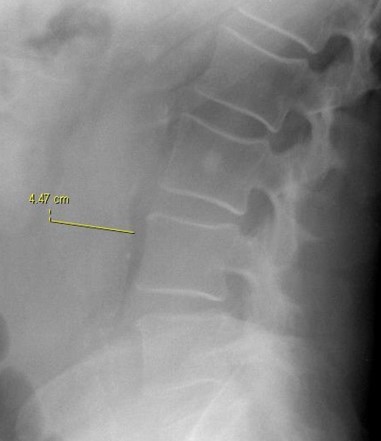
Lateral lumbar spine view
The sagittal dimension of the abdominal aorta measures 4.4 cm. well beyond the accepted normal measurement of 3-3.5 cm. (1)
The radiodensity seen superimposed the VB is in soft tissue.
The transverse dimension of the abdominal aorta measures 4. 4 cm. (N=3.0-3.5 cm). <sub> (1)</sub>
The radiodensity superimposed on the L3 VB is within soft tissue as are the densities anterior to the upper lumbar VB.
A CT scan of the abdomen was ordered.
AAA:
- most common cause of death in Western world.
- atherosclerotic etiology is most common.
- US can be used for AAA screening & following small dilations.
- CT angiography is considered gold standard.
- > 5.0 mm enlargement within 6 month span warrants intervention. >sub>(3)</sub>
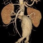
A 3D reformatted model of an abdominal aortic aneurysm is in its common infra-renal location. <sub> (2)</sub>
DISCUSSION: The mass effect of the OPLL seen on the axial CT image may causes stages of cord damage described by Radiopaedia.com seen above. The cases of OPLL that occupy 30-60% of the cross-sectional diameter are more likely to show myelopathy. Laterally deviated OPLL masses have a higher risk of myelopathy than those at the midline (5)
REFERENCES:
- Essentials of Skeletal Radiology, Yochum & Rowe 2nd. Ed. p. 1309
- From the case rID: 8190
- https://radiopaedia.org/articles/abdominal-aortic-aneurysm