50 yo/M patient presents with non-specific neck discomfort.
No prior trauma, surgeries, carcinoma orcorticosteroid/opioid regimen reported.
What do you see?
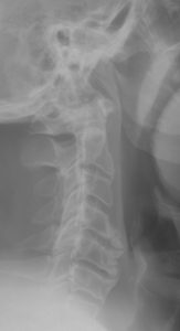
Cervical spine- lateral
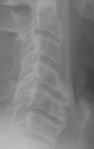
Cervical spine- close up exam.
The C2 disc shows annular calcification. Spondylosis/DISH like change is seen at C4-5 motor unit with degenerative disc disease at C5.
The keys finding is the thin vertical density posterior to the C2-4 levels involving posterior longitudinal ligament. This is best seen on the close -up view.
Stages of spinal cord damage by OPLL
- stage 0: normal or mild compression of the anterior horn without neuronal loss
- stage 1: mild compression of the anterior horn with partial neuronal loss
- stage 2: marked deformity of anterior horn; severe neuronal loss
- stage 3: severe spinal cord damage
T2-weighted sequences are considered the most effective in the evaluation of spinal cord compression. (1) Radiopaedia.com
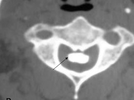
The axial CT scan – bone window shows OPLL which causes mass effect on the cord. (4) mrimedicvn.blogspot
DISCUSSION: The mass effect of the OPLL seen on the axial CT image may causes stages of cord damage described by Radiopaedia.com seen above. The cases of OPLL that occupy 30-60% of the cross-sectional diameter are more likely to show myelopathy. Laterally deviated OPLL masses have a higher risk of myelopathy than those at the midline (5)
REFERENCES:
- https://radiopaedia.org/articles/ossification-of-the-posterior-longitudinal-ligament
- https://bmcmusculoskeletdisord.biomedcentral.com/track/pdf/10.1186/s12891-018-2227-z.pdf
- Mader R, Baraliakos X,
Eshed I, et al. Imaging of DISH). RMD Open 2020;6:e001151. doi:10.1136/ rmdopen-2019-001151 - http://mrimedicvn.blogspot.com/2011/04/posterior-spinal-ligaments-ossification.html
- https://radsource.us/ossification-posterior-longitudinal-ligament-bone-bad-place/