11 yo/female patient softball player present with left groin discomfort.
No prior trauma, surgeries, carcinoma or corticosteroid regimen.
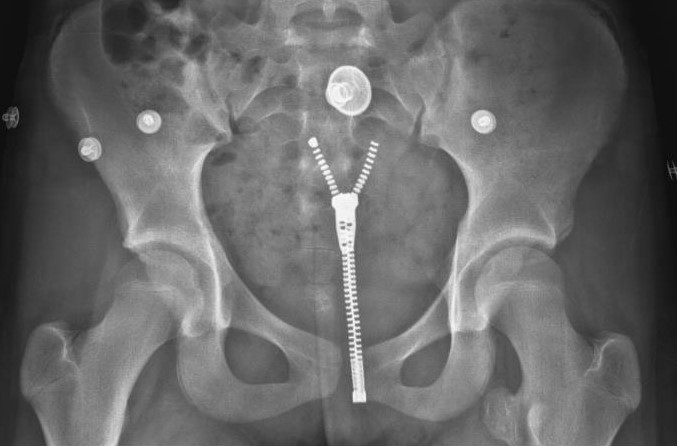
Medial & slightly superior to the left lesser trochanter, a 1. 7 x 2.0 mm. exostosis is seen with cortices continuous with the parent. Matrix calcification ( multiple radiodense dots) are present. A CT scan was ordered for optimal radiographic evaluation. The coronal bone CT scan allows accurate anatomic locationalization of the suspected osteochondroma and its 3D planar capability also allows precise correlation with the neurovascular bundle.
Radiopaedia.org diagram illustrated the relationship of the circumflex branches of the femoral artery to the lesser trochanter. (1) The block dot represents entry point for femoral artery cardiac catherization.
A suspected osteochondroma can be evaluated with a non-contrast CT scan of the pelvis (to include the hip joints)(2)
Malignant transformation of a single osteochroma is low. A cartilage cap thickness > 1.5 cm. (on advanced imaging) should be a suspicious finding for malignant transformation. (2)