40 year old female presents to a DC office complaining of non-specific lower discomfort. No prior history of trauma, surgery, carcinoma or corticosteroid/opioid regimen.
40 year old female presents to a DC office complaining of non-specific lower discomfort. No prior history of trauma, surgery, carcinoma or corticosteroid/opioid regimen.
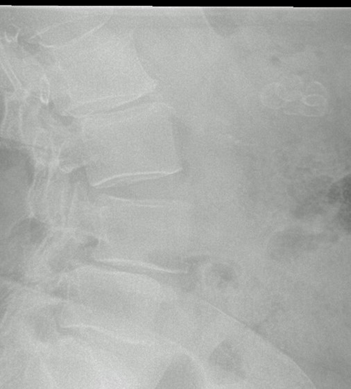
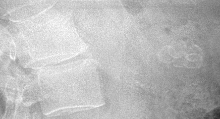
Fig. 1 AP view – notice pendulous abdomen&subtle ring-enhanced radiolucencies of the RUQ.
FIG.2 Lateral view – shows L2 spondylosis, L4 degenerative anterolisthesis. L5 disc narrowing with retrolisthesis. Ring-enhanced radiolucencies -intraperitoneal in location.
FIG.3 – CLOSE UP VIEW – Showing multiple (5) cholelisthiasis. Diagnostic US of the abdomen is warranted.
FIG.4- Transverse scan of GB with stones and vertical radiolucencies (acoustical shadowing). Blue elliptical region = GB.
FINDINGS:
In the RUQ, multiple ring -enhanced radiolucencies are present – all intraperitoneal in location.
Spinal changes include: L4 degenerative spondylolisthesis, L5 disc narrowing, mild SI disease.
IMPRESSIONS:
1. CHOLELITHIASIS – RUQ
2. L2 SPONDYLOSIS WITH RETROLISTHESIS.
3. L4 L4 DEGENERATIVE SPONDYLOLITHESIS.
4. L5 DISC NARROWING.
Only 15%–20% of gallstones have enough calcium within to be visible on plain x-rays therefore radiographs underestimates the number of stones present. (3) This necessitates a follow up examination with diagnostic ultrasound examination of the abdomen.
At ProImaging we provide expert chiropractic radiology interpretation.

